Fundus Photography
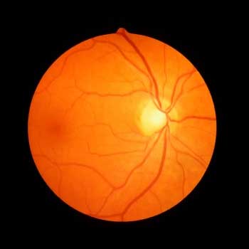
Fundus photography involves capturing a photograph of the back of the eye i.e. fundus. Our specialized fundus cameras consist of an intricate microscope attached to a flash enabled camera and are used in fundus photography. The main structures that can be visualized on a fundus photo are the central and peripheral retina, optic disc and macula. We perform fundus photography with colored filters, or with specialized dyes including fluorescein and indocyanine green. Our treatment starts as soon as the patient sits at the fundus camera with their chin in a chin rest and their forehead against the bar. Our ophthalmic photographer focuses and aligns the fundus camera. A flash fires as our photographer presses the shutter release, creating a fundus photograph. Our ophthalmologists use these retinal photographs to follow, diagnose, and treat eye diseases.